
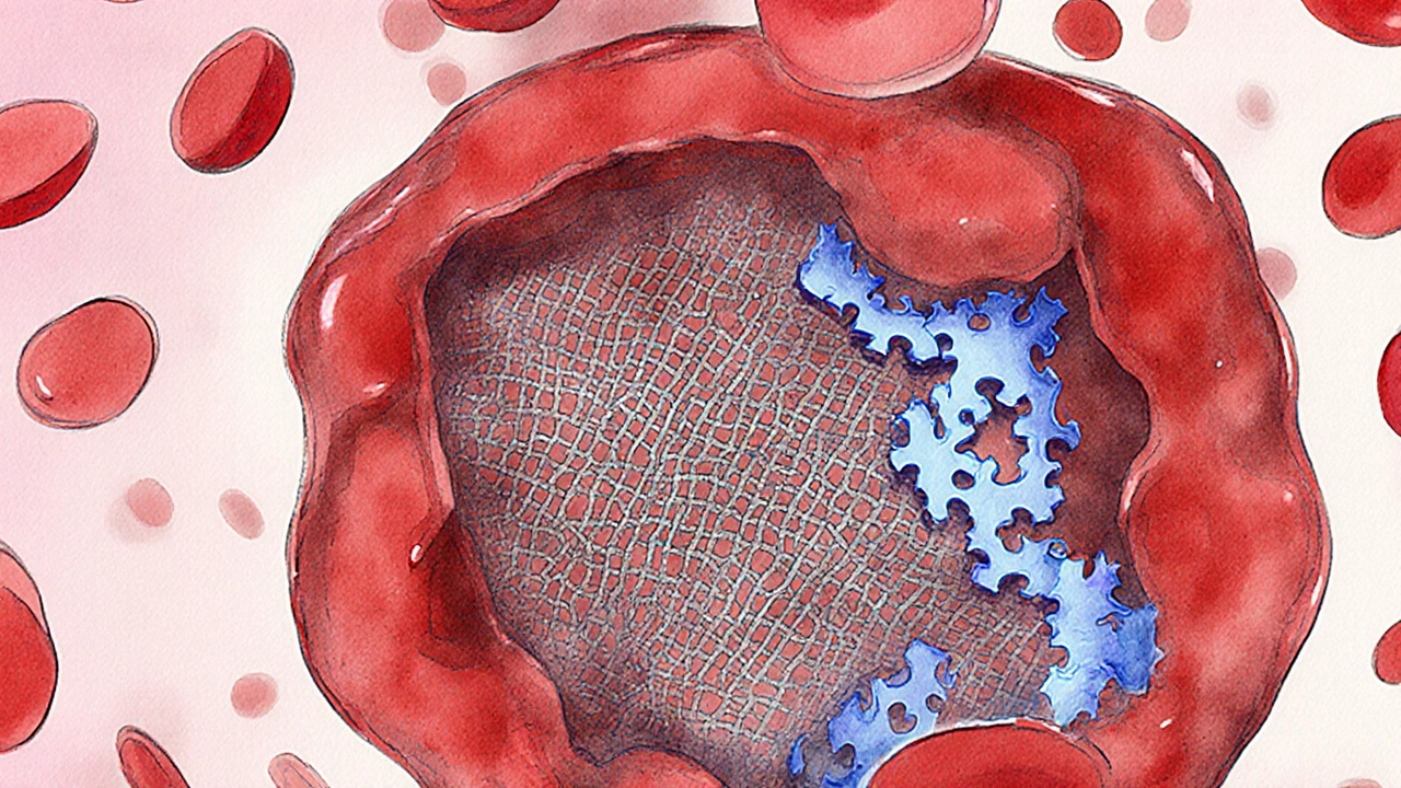
Hemophilia is a genetic bleeding disorder caused by deficiency of clotting factor VIII or IX. von Willebrand disease is the most common inherited bleeding disorder, stemming from a deficit or dysfunction of von Willebrand factor. Factor XI deficiency is a rare autosomal recessive condition that reduces factor XI activity, leading to delayed bleeding. Platelet function disorders are a group of conditions where platelets fail to aggregate properly despite normal clotting factor levels. Coagulation cascade is the series of enzymatic reactions that convert fibrinogen to fibrin, forming a stable clot. Clotting factor replacement therapy is the standard treatment that supplies missing factors intravenously to stop or prevent bleeding. Gene therapy is a cutting‑edge approach that introduces functional copies of defective genes into patients’ cells. Diagnostic tests such as prothrombin time (PT) and activated partial thromboplastin time (aPTT) help pinpoint which part of the cascade is impaired. Understanding how these pieces fit together clears up the confusion many feel when they hear “bleeding disorder”. Below, we break down the main categories, point out what sets hemophilia apart, and give you a quick roadmap for talking to doctors.
Quick Take
- Hemophilia involves factor VIII (type A) or IX (type B) deficiency; most other rare bleeding disorders affect different factors or platelets.
- Inheritance patterns differ: hemophilia is X‑linked, while many rare disorders are autosomal recessive or dominant.
- Symptoms overlap (joint bleeds, bruising) but onset, severity, and trigger events vary.
- Diagnosis relies on specific lab panels: aPTT is prolonged in hemophilia, PT stays normal.
- Treatment options range from on‑demand factor replacement to long‑term gene therapy.
How Hemophilia Is Defined
People with hemophilia typically experience spontaneous bleeds into joints and muscles, especially knees, elbows, and ankles. The condition is classified by severity:
- Severe: less than 1% normal factor activity - bleeds can occur without injury.
- Moderate: 1‑5% activity - bleeding usually follows minor traumas.
- Mild: 5‑40% activity - symptoms appear only after significant injury or surgery.
Because the defective gene sits on the X chromosome, almost all severe cases are male; females are usually carriers, though rare lyonization can produce symptomatic women.
Other Rare Bleeding Disorders at a Glance
Beyond hemophilia, clinicians encounter a handful of conditions that share the “bleeding” label but differ in cause and management.
| Disorder | Inheritance | Deficient Factor / Mechanism | Typical Lab Finding | Common Clinical Features |
|---|---|---|---|---|
| Hemophilia A | X‑linked recessive | Factor VIII | Prolonged aPTT, normal PT | Joint & muscle bleeds, prolonged bleeding after trauma |
| Hemophilia B | X‑linked recessive | Factor IX | Prolonged aPTT, normal PT | Similar to Hemophilia A, slightly milder in some cases |
| von Willebrand disease | Autosomal dominant (most types) | von Willebrand factor (carrier of factor VIII) | Prolonged bleeding time, reduced ristocetin co‑factor activity | Nosebleeds, mucosal bleeding, menorrhagia |
| Factor XI deficiency | Autosomal recessive | Factor XI | Prolonged aPTT, normal PT | Delayed postoperative bleeding, mild joint bleeds |
| Platelet function disorder (e.g., Glanzmann thrombasthenia) | Autosomal recessive | Defective GPIIb/IIIa receptor | Normal PT/aPTT, prolonged bleeding time | Pinpoint bruising, mucosal bleeding, poor wound healing |
Why Lab Tests Matter
When a patient presents with unexplained bruising, a doctor orders a panel that typically includes PT, aPTT, bleeding time, and specific factor assays. The pattern tells us which part of the Coagulation cascade is broken.
For hemophilia, the aPTT is the star player-it's prolonged because the intrinsic pathway (where factor VIII and IX operate) is slowed. In contrast, von Willebrand disease shows a normal aPTT but an abnormal ristocetin co‑factor activity, pointing to the carrier protein rather than the cascade itself.
Treatment Landscape: From Replacement to Gene Therapy
Traditional Clotting factor replacement therapy involves infusing plasma‑derived or recombinant factor concentrates on demand or as prophylaxis. Regular prophylaxis, especially in severe hemophilia, reduces joint damage by 80% compared with on‑demand treatment.
Newer options include:
- Extended half‑life products: PEGylated or Fc‑fusion factors stay in the bloodstream longer, cutting infusion frequency.
- Emicizumab: A bispecific antibody that mimics factor VIII activity, useful for patients with inhibitors.
- Gene therapy: A single infusion of an adeno‑associated virus (AAV) vector delivers a functional copy of the deficient gene. Early‑phase trials in hemophilia A show factor VIII levels >50% in 70% of participants, effectively turning severe disease into a mild form.
Rare disorders like Factor XI deficiency often respond to fresh frozen plasma or specific factor XI concentrates, but because bleeding is less frequent, prophylaxis is rarely used.
Living With a Bleeding Disorder: Practical Tips
Whether you have hemophilia or a different rare condition, daily habits can make a big difference.
- Know your numbers: Keep a written record of your factor levels, inhibitor status, and medication schedule.
- Communicate with caregivers: Share your diagnosis, recommended treatments, and emergency plan with teachers, coaches, and employers.
- Protect joints: Low‑impact activities like swimming or cycling reduce the risk of joint bleeds compared with high‑impact sports.
- Watch for early signs: Swelling, warmth, or reduced range of motion in a joint may signal a bleed that needs prompt factor infusion.
- Stay up‑to‑date on therapies: Clinical trials for gene therapy and novel agents are expanding; discuss eligibility with your hematologist.
When to Seek Specialist Care
General practitioners can manage mild cases, but severe hemophilia or any rare bleeding disorder benefits from a multidisciplinary hemophilia treatment center. These centers offer:
- Comprehensive coagulation labs that run factor assays on the same day.
- Physical therapy programs designed to protect joints.
- Psychosocial support for patients and families dealing with chronic illness.
- Access to clinical trials and emerging therapies.
Early referral improves outcomes and lowers long‑term healthcare costs.
Frequently Asked Questions
How is hemophilia different from von Willebrand disease?
Hemophilia is caused by a missing clotting factor (VIII or IX) affecting the intrinsic pathway, while von Willebrand disease involves a faulty carrier protein that also stabilizes factor VIII. Lab tests show a prolonged aPTT in hemophilia but a normal aPTT and abnormal ristocetin co‑factor activity in von Willebrand disease.
Can females have hemophilia?
Because the gene is on the X chromosome, most females are carriers. Rarely, due to skewed X‑inactivation, a woman can show mild to moderate symptoms.
What are the newest treatments for severe hemophilia?
Extended‑half‑life factor concentrates, bispecific antibodies like emicizumab, and AAV‑based gene therapy are leading the field. Gene therapy can raise factor levels to mild‑disease range after a single infusion.
Why do some rare bleeding disorders cause delayed bleeding?
Disorders such as Factor XI deficiency affect the later stages of the intrinsic pathway, so clot formation starts but takes longer to stabilize, leading to bleeding that appears hours after injury or surgery.
Is prophylactic treatment necessary for all rare bleeding disorders?
Prophylaxis is standard for severe hemophilia because joint bleeds are frequent. For many other rare disorders, bleeding episodes are sporadic, so on‑demand therapy is usually sufficient.
Kevin Ouellette
September 29, 2025 AT 05:24Just found out my nephew has hemophilia A last month 😢 This post broke it down so clearly-I finally get why his aPTT is high but PT is normal. Thanks for the clarity! 🙌
Tanya Willey
September 29, 2025 AT 16:46Wait… so you’re telling me this whole ‘gene therapy’ thing is just Big Pharma’s way to lock us into lifelong dependency? They’ve been hiding the truth about clotting factors since the 80s. Remember the tainted blood scandal? 🤔
sarat babu
September 30, 2025 AT 14:13Brooooooo-this is LIFE-CHANGING info!! 🤯 Hemophilia isn’t just ‘bleeding’-it’s a whole SYSTEM failure!! And don’t even get me started on how von Willebrand’s is being downplayed?? They’re treating it like a nosebleed issue when it’s a FULL BLOOD CRISES!!! 😭
Wiley William
September 30, 2025 AT 14:22Yeah right, gene therapy? That’s just a placebo wrapped in biotech jargon. They’ve been saying this for 20 years. Where’s the long-term data? Who funded these trials? I’ve seen this movie before-remember the ‘miracle cure’ for MS? It vanished overnight.
Richard H. Martin
October 1, 2025 AT 13:06Our country has the best medical system in the world-why are we even talking about this? In America, we don’t have ‘rare bleeding disorders’-we have patients who get treated FAST. If you’re not getting care, you’re doing it wrong.
Tim H
October 1, 2025 AT 21:25so i had this friend who had like factor xi deficiency and he said he only bled after surgery but like… why does the aPTT go up but not the PT? i always thought they were the same thing?? also i think the table is wrong because my cousin’s lab results were different??
Umesh Sukhwani
October 2, 2025 AT 02:48Thank you for this meticulously researched and clinically accurate overview. The distinction between intrinsic pathway defects and platelet dysfunction is often misunderstood even among medical students. The emphasis on multidisciplinary care centers is particularly vital in resource-limited settings. May this guide reach those who need it most.
Vishnupriya Srivastava
October 2, 2025 AT 07:34Interesting how they frame gene therapy as ‘cutting-edge’ when it’s still experimental. The real issue is access-most patients in developing countries can’t even get factor concentrates, let alone gene therapy. This reads like a marketing brochure for biotech.
Matt Renner
October 2, 2025 AT 15:05The diagnostic algorithm described here is textbook. However, I would add that bleeding time assays are largely obsolete in modern labs. Most centers now rely on platelet function analyzers (PFA-100/200) and VWF antigen/activity ratios for von Willebrand disease. The table should be updated accordingly.
Wayne Rendall
October 3, 2025 AT 06:41Excellent breakdown. The distinction between X-linked recessive and autosomal inheritance patterns is crucial for genetic counseling. I would only suggest clarifying that ‘mild’ hemophilia isn’t benign-many patients still develop chronic arthropathy without prophylaxis.
Ifeoluwa James Falola
October 3, 2025 AT 11:43This is helpful. Many don’t realize that Factor XI deficiency can cause bleeding after dental work or childbirth. Not everyone bleeds right away. Delayed bleeding is real.
Adam Phillips
October 4, 2025 AT 03:04What is a clot really? Is it just fibrin or is it a living thing? Are we treating the symptom or the soul of the problem? We call it a disorder but maybe it’s just nature adjusting…
Julie Lamb
October 4, 2025 AT 10:36I’m so glad someone finally explained this in a way that doesn’t make me feel like a burden. My daughter has vWD and I used to think she was just ‘clumsy’… now I know how to protect her. Thank you 🤍
april kakoske
October 5, 2025 AT 01:50the fact that we can fix genes now… it’s wild. like we’re not just treating bodies anymore we’re rewriting them. feels like sci fi but also kind of beautiful
Pradeep Meena
October 5, 2025 AT 15:36Why do we waste money on gene therapy when we can just give people more blood? In India we fix everything with medicine and willpower. These Americans think they need fancy machines to bleed properly
Rishabh Jaiswal
October 6, 2025 AT 11:14wait so factor XI is autosomal recessive? i thought it was dominant? and aPTT is prolonged in all of them right? i think this article is wrong because my cousin’s doctor said something different…
May Zone skelah
October 6, 2025 AT 22:47Let’s be real-this isn’t just about clotting factors. It’s about the existential loneliness of living with a body that betrays you. The silent screams in the ER when no one understands why you’re bleeding… the guilt of being a ‘burden’… the way your mother’s eyes fill with fear every time you fall. This article? It’s a surface skim. The real story is written in the quiet tears at 3 a.m.
Dale Yu
October 7, 2025 AT 11:15They say gene therapy is the future but they never tell you the cost. You think your insurance will cover it? Nah. You’re gonna be stuck with $2 million bills and a lifetime of waiting for the next trial. This is capitalism with a stethoscope
Kshitij Nim
October 8, 2025 AT 04:18Great summary. One thing missing: the importance of physical therapy. Many patients focus only on factor infusions but joint protection is half the battle. Stretch, strengthen, swim. Don’t wait for the bleed to start moving.
Scott Horvath
October 8, 2025 AT 11:59so i just found out my kid has mild hemophilia and honestly i thought it was just a ‘minor thing’… but now i realize it’s like carrying a time bomb in your knees. this post changed everything. thank you for not making me feel dumb for not knowing